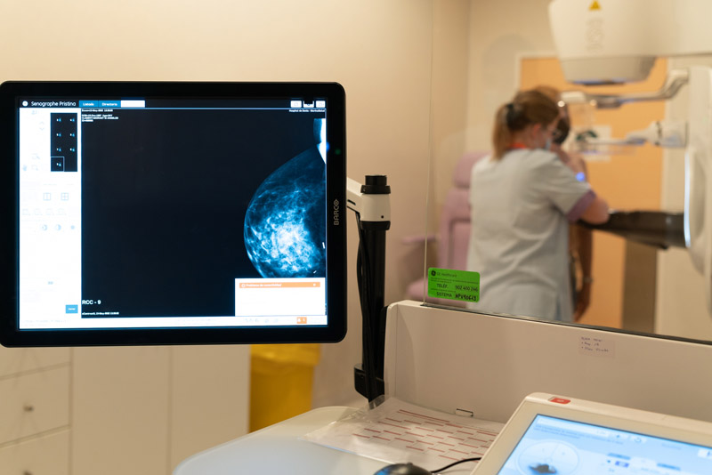
- The precision and capability of this technology is so high that it is used in the check-ups of 100% of patients that have overcome breast cancer and also for those that are unable to have an MRI
- The corporate manager of the healthcare group’s Breast Department takes a very positive view of these diagnostic tests, after more than two years of carrying them out in the university hospitals of Torrejón and Vinalopó (Elche)
Detecting a tumor when it has barely begun to form. Contrast-enhanced mammography at the Ribera healthcare group‘s hospitals is so precise that it enables them to detect the network of vessels that are formed at the start of a tumor. In this way, specialists with extensive experience in breast pathologies, such as Doctor Julia Camps, the corporate manager of the Breast Department, can diagnose the formation of a tumor even when it has barely begun to grow, and thus make clinical decisions and a therapy plan for the patient earlier. “Early detection is key in order to achieve the best results in the treatment of each patient”, affirms Doctor Camps.
“Contrast-enhanced mammography is a very new technique, which consists of injecting an iodine-based contrast into the patient and bringing to light tumors that a normal mammogram would not detect because structures overlap and many tumors are hidden”, explains Julia Camps.
After two and a half years of applying this diagnostic technique at Torrejón University Hospital, and just over two years at Vinalopó University Hospital, the results of the use of the contrast-enhanced mammography are very good, especially in cases whereby patients had breast cancer previously, as well as in those that where unable to have an MRI”, explains Doctor Camps, referring to women with heart problems, claustrophobia or those who are overweight.
Contrast-enhanced mammography at these two hospitals within the Ribera healthcare group is up to 30% more sensitive than a conventional mammogram, and is able to detect tumors of just 4 millimeters, and even the formation of vessels at the beginning, which normally, she adds, “wouldn’t show up until two years later”. In the case of patients that have previously had breast cancer, it is sometimes difficult to small tumors with a conventional mammogram, as the corporate manager of the Breast Department explains, “due to the changes in the breast after the previous treatment”. In addition, she adds, “the morphology or form of these lesions may go unnoticed if the surrounding tissue is very dense or heterogeneous and, for that reason, we take advantage of the capacity of functional techniques such as contrast-enhanced mammography or MRIs to highlight the tumoral angiogenesis or function that allows for the detection of cancer independently from its morphology”. With this technology, she assures, “we are in a position to give patients maximum guarantees that a new lesion has not appeared, no matter how small, when they have their follow-up mammogram”.
Doctor Camps reiterates the “vital importance” of these contrast-enhanced mammograms for patients that, for various reasons, are unable to have an MRI. “Patients that have claustrophobia, that cannot lie on their front or that have cardiac or respiratory problems, can have access to their tumoral map thanks to this technology, and with the same reliability as that of an MRI”, she affirms.
The surgical coordinator of the Breast Department, Doctor Lorenzo Rabadán, is of the same opinion. “New technology, such as MRIs with diffusion software and contrast-enhanced mammograms offer us an image of breast cancer that we have never had before. You could almost say that we operated based on hearsay, because we didn’t see the full reality of the tumor, and this technology now brings us closer to the reality of their tumor, with its appendages and nodules that we didn’t see earlier”, she explains.
In this video, Doctor Julia Camps, Doctor Laia Bernet and Doctor Lorenzo Rabadán explain what this is and how a contrast-enhanced mammogram is done.


Recent Comments